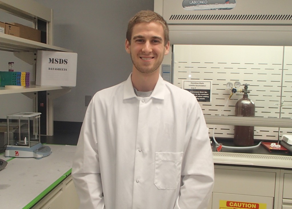
Greening in Dr. Muldoon’s Laboratory at the Engineering Research Center
Welcome to my blog! Let me begin by introducing myself a bit more. I’m a senior biomedical engineering major in the Honors College here at the University of Arkansas. In addition to my academics, I work in Dr. Muldoon’s research laboratory. I’ve been in college for three full years now, and I know people say this all the time, but the time has flown by. I’ve been lucky to have the support of my family and friends, who keep me level headed in times of stress that come along with college.
I began my undergraduate career with only a vague conception of what college really meant. I wanted to make something of myself, to challenge myself, but I didn’t know how. I’ve always had a diverse set of interests, including the life sciences, math, athletics, and art, especially sketching and filmmaking. But how do I combine these passions to enrich my college (and life) experience? This became my goal. In the back of my mind was something my dad had always taught me…live life by taking care of yourself first, both mentally and physically. Only by enriching yourself can you share your passions and abilities with the world and make it a better place to live. So I set out on my own personal adventure…an adventure I’m still on.
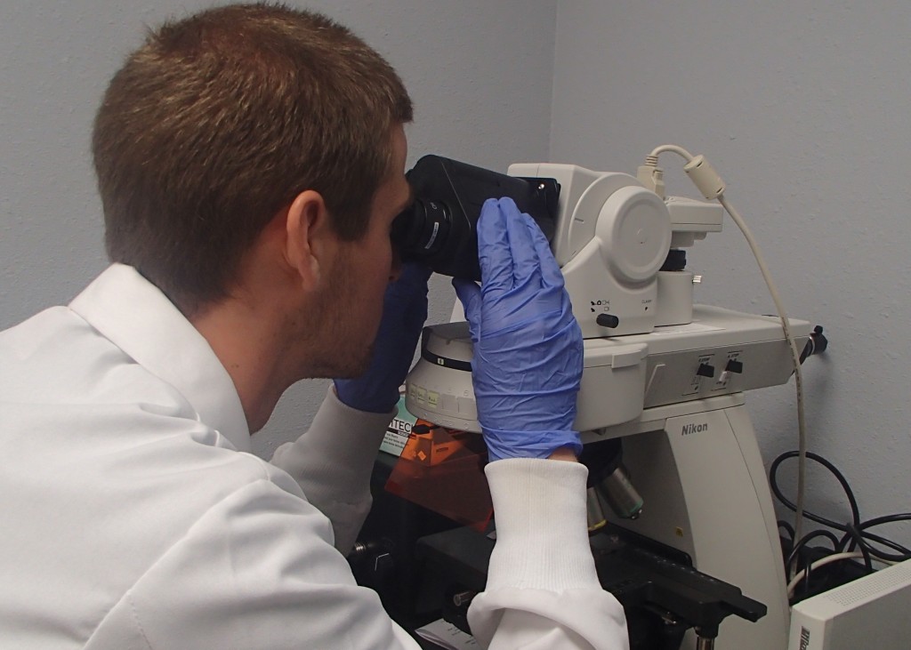
Greening gathering images using a microscope.
It was the beginning of my junior year that the biomedical engineering major was created. Along with this new major came the addition of several new biomedical engineering faculty members, including Dr. Timothy Muldoon, the principal investigator of the Translational Biophotonics and Imaging Laboratory. Now this is quite the impressive name, but put simply, the lab uses image analysis to diagnose disease, primarily cancer. It’s the combination of art and science in perfect harmony. Because of that, I knew this lab would be an excellent place to conduct research for my honors thesis. Throughout my junior year, I performed preliminary research on the diffuse reflective microendoscope project, an innovative new approach for the early diagnosis of oral cancer. My efforts paid off when I was awarded an Honors College Undergraduate Research Grant for the 2013-2014 school year. I decided to stay in Fayetteville this past summer to perform more preliminary work on this project.
Specifically, my research is focused on the development of a non-invasive diffuse reflective microendoscope, henceforth referred to as DRME, which could improve the early detection of cancer in epithelium, such as the lining of the oral cavity or gastrointestinal tract. Improvement in early detection methods should significantly increase survival rates. In practice, an illumination fiber will deliver infrared light to a small area of epithelial tissue, and a camera will capture the light that scatters back out of the tissue. The amount of light that scatters back out, called diffusely reflectance, is critical in determining whether abnormalities exist beneath the surface of the epithelium. Cancerous and precancerous tissue will scatter back less light than normal, healthy tissue. Therefore, the goal of the DRME is to provide clinicians with information about abnormalities that exist beneath the visible surface of epithelial tissue, especially in the oral cavity.
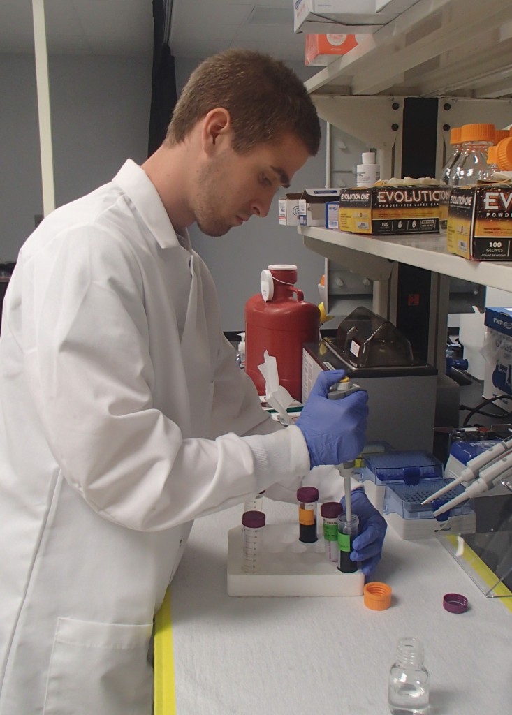
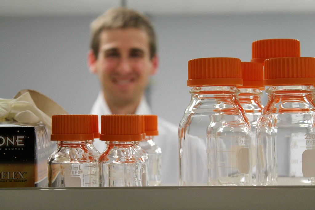
Greening creating tissue phantoms
As of now, my research can be divided into two goals. The first goal is to create optical tissue phantoms. Because this is a new approach to cancer detection, we cannot jump right in to testing our imaging system on human subjects. Therefore, I will be creating “phantoms,” thin gel-like layers that will replicate the optical properties of epithelial tissue and will provide a means of verification for the DRME. These optical phantoms will be designed to have several different layers with distinct optical properties by using materials with known absorbing and scattering properties. Currently, I’m designing a procedure that will allow me to build phantoms with specific depths (down to a tenth of a millimeter!) to replicate the different layers in actual epithelial tissue.
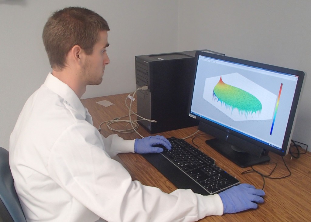
Greening running a Monte Carlo simulation
This brings me to the second goal of my research, to run computer simulations as a means of modeling light transport through epithelial tissue. When light enters tissue, it penetrates the surface, bounces around within, and then bounces back out (diffuse reflectance). This computer model, called a Monte Carlo simulation, uses probability to predict how light behaves within tissue. I can modify the Monte Carlo code based on the properties of actual tissue or optical phantoms, such as scattering and absorbing values. Once I’ve fine-tuned the computer simulation, we’ll be able to make final adjustments to the DRME imaging system. These final adjustments will ensure maximum light delivery to the optical phantoms, so that our data is reliable and reproducible. Then we’ll be one step closer to testing the DRME on human tissue, although this is still a long way off.
I’m excited to see where my research will take me these next two semesters. I’ll be updating my research blog as often as possible, so stick around with me this year to see all the craziness that comes along with the roller coaster of success, failure, and learning in the world of biomedical research!
Greening conducting research in the Translational Biophotonics and Imaging Laboratory