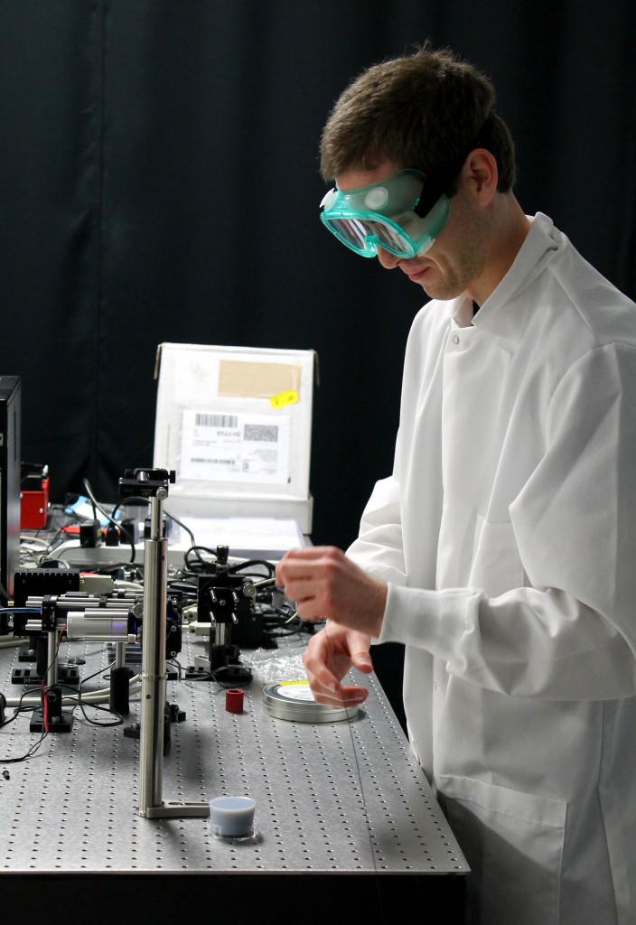
Gage turning on the diffuse reflective microendoscope
Welcome to my second blog entry! During the fall 2013 semester, I wrote about my research experience thus far as part of the College of Engineering Honors Program. I am now in my last semester before I graduate and am in the process of writing my honors thesis.
My research is focused on designing a diffuse reflective microendoscope (DRME), a non-invasive method that may aid in the early detection of cancer in the oral cavity or gastrointestinal tract. Under this umbrella, my research is divided into two goals. My first goal is to develop a method for creating optical phantoms that replicate the optical properties of epithelial tissue. My second goal is to build the DRME and computationally model how light behaves through phantoms and epithelial tissue.
The majority of my research last semester was the development of my first goal. I was able to design ultra-thin phantoms that replicate optical properties of human epithelial tissue, such as in the oral cavity. When epithelial tissue becomes cancerous, it becomes more optically absorbing, meaning that cancerous lesions appear darker than surrounding healthy tissue. While a dark cancerous region on the tissue surface is easily detectable by normal endoscopic methods, occasionally cancer will exist below the tissue surface. This results in undetected cancer and abnormalities. Thus, the DRME attempts to reduce the amount of false negatives. My goal is to detect hidden cancers that are not easily detectable by conventional clinical methods. Because of this, I created “dark” phantoms to simulate cancerous tissue and “light” phantoms to simulate healthy tissue. Then, I was able to stack individual layers to create a multi-layered phantom with “dark” cancerous regions buried at varying depths within “light” healthy regions.

Optical Phantom with dark “cancerous” regions and light “healthy” regions
The multi-layered phantom will allow me to test the DRME before actual animal and human trials begin. However, in order to begin testing, we first had to build the DRME. Along with my faculty mentor, Dr. Muldoon and fellow classmate, Josh Hutcheson, we built the DRME in January of 2014. This was one of the more exciting and challenging experiences as an undergraduate researcher. The final construction involved several building and rebuilding processes. In fact, it will continue undergoing small modifications for some time. The primary construction challenges arise because of “dual-channel” aspect of the DRME.
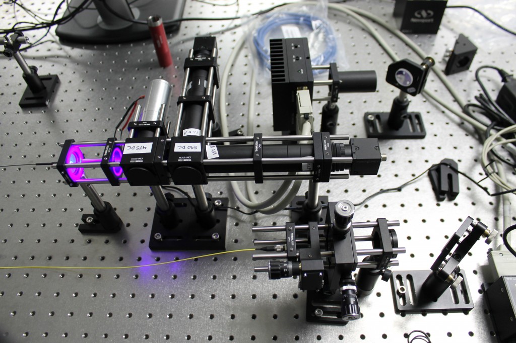
Diffuse Reflectance Microendoscope (DRME) with perpendicular channels
The DRME has two separate channels. The first channel will detect cellular structure on the surface of the oral cavity. However, as mentioned previously, not all oral cancer exists on the cell surface. Therefore, the second channel will detect sub-surface “diffuse reflectance,” which can detect abnormalities within deep tissue. In our initial construction process, we built the channels perpendicular to each other. Our hopes were that each camera, attached to the end of each channel, would be detecting the exact same area. We discovered this was not the case. While the cameras were detecting areas close to one another, one camera was slightly out of alignment. Imagine trying to take two pictures of the same object. Most likely, although the images will be very similar, they will not be exactly the same. In one image, the object may be shifted ever so slightly. This was the problem we were experiencing. We needed the cameras to take pictures of the exact same space at the same time. To correct this problem, we disassembled our entire system. In “version 2” of the DRME, we made each channel parallel to one another, instead of being at right angles. This was possible by the addition of a mirror in the middle of the second channel. The mirror is adjustable, so we can tune the incoming light rays to hit the exact same spot on the second camera as they do in the first camera.
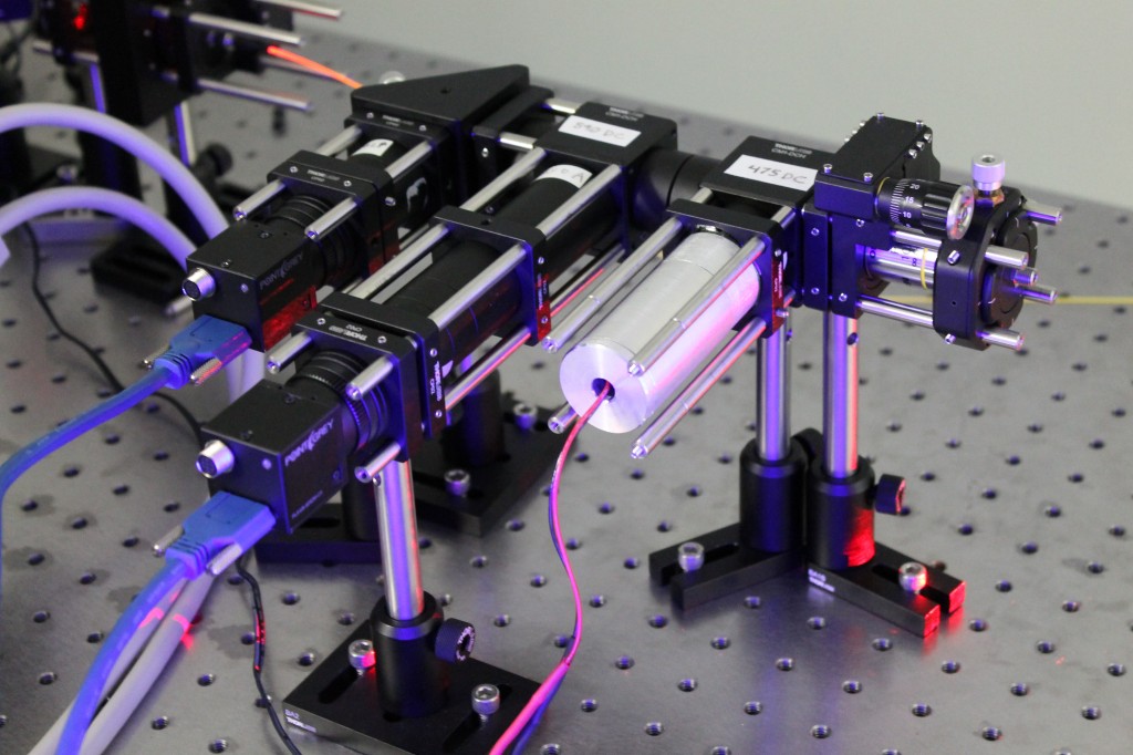
Diffuse Reflectance Microendoscope (DRME) with parallel channels
While the DRME has been built and is currently undergoing validation with optical phantoms, there are still many steps to be taken. I’ll be keeping you updated on future developments!
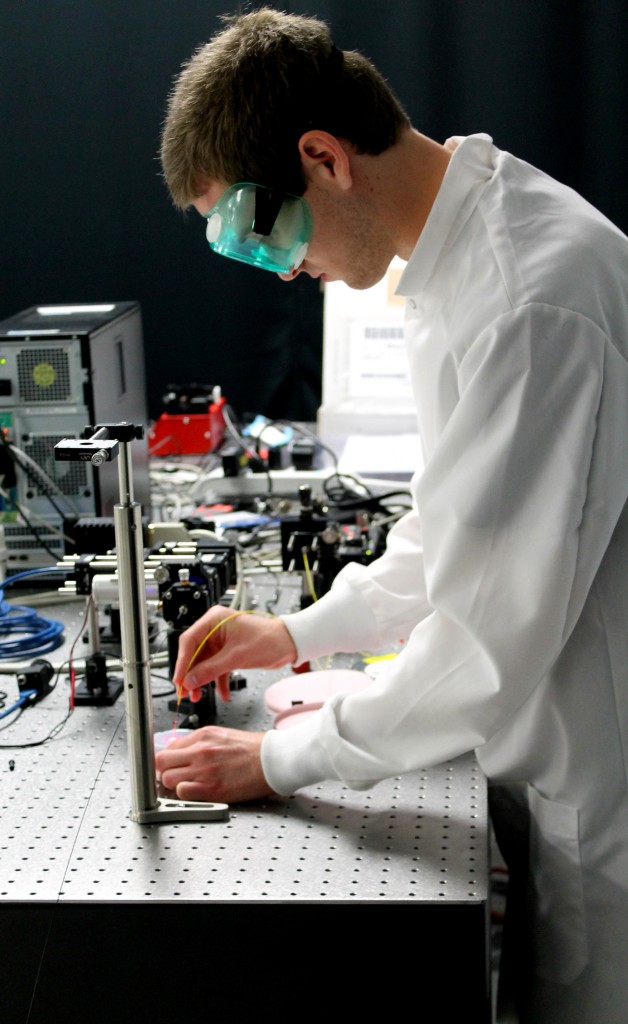
Gage shining red light onto a multi-layered phantom